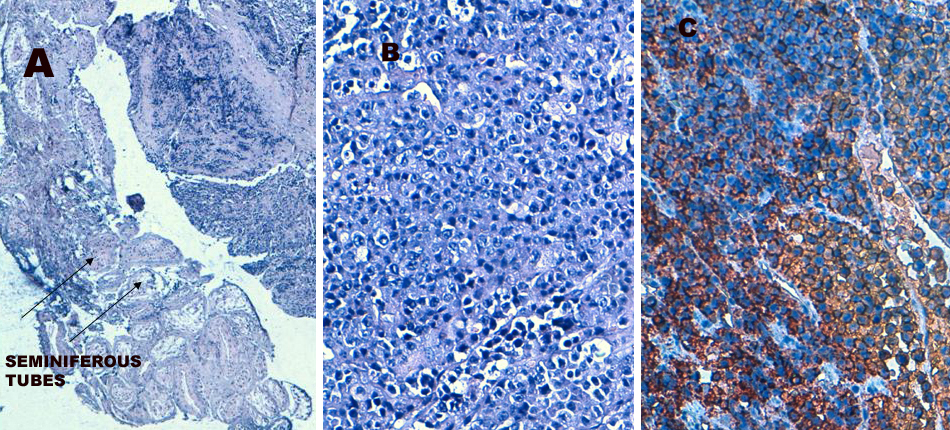
Figure 1. Plasma cell proliferation involving testicular parenchyma (A). Plasmocytoid cells (B). Tumor cells intensively marked by the anti-CD138 antibody on immunohistochemistry (C).